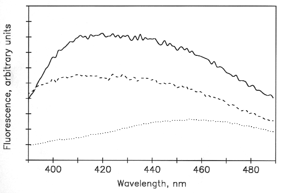


Emission spectrum of 2-AB-taxol in the presence and absence of the
colchicine-tubulin complex. The colchicine-tubulin complex was formed
by incubating tubulin with a 10-fold excess of colchicine for 30
minutes at 37 degC prior to removal of unbound ligand
by rapid gel filtration. Dotted curve: 2-AB-taxol
 2 (M) in PME buffer.
Solid curve: 2
  M 2-AB-taxol
and 5
  M tubulin-colchicine complex in
PME buffer were incubated for 20 min at 25 degC prior to
acquisition of the spectrum. Dashed curve:
2
  M 2-AB-taxol and
5
  M tubulin-colchicine complex in PME buffer
were incubated for 20 min at 37 degC prior to
addition of 10
  M taxol.
The excitation wavelength was 320 nm. Note the decrease in intensity of
the dashed curve relative to the solid curve. Electron micrographs of the
solutions used for the solid and dashed curves showed no high order tubulin
structures.
